

Your heart is a living, moving organ — and a standard ECG, while valuable, only captures its electrical activity. To truly see your heart in action — its walls contracting, its valves opening and closing, its chambers filling and emptying — doctors rely on the 2D echocardiography test. This remarkable imaging investigation gives cardiologists a real-time window into the mechanical function of your heart, making it one of the most important diagnostic tools in modern cardiology.
Whether your doctor has recommended a 2D echocardiography test after an abnormal ECG, or you are here to understand what this test involves before your appointment, this complete guide by Dr. T. Sandeep — Interventional Cardiologist at Jayam Hrudayalaya — answers every question you might have.
What is a 2D Echocardiography Test?
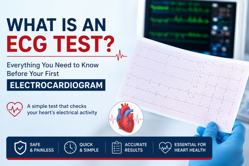
A 2D echocardiography test is a cardiac ultrasound examination that uses high-frequency sound waves to create real-time, two-dimensional moving images of your heart. The term “echo” is short for echocardiography — a combination of the words “echo” (sound reflection) and “cardiography” (heart recording).
Unlike an X-ray that shows bones and static structures, or an ECG that reads electrical signals, the 2D echocardiography test produces live video images of your heart while it is beating. This allows your cardiologist to directly observe:
- The four chambers of the heart (left atrium, right atrium, left ventricle, right ventricle)
- All four heart valves (mitral, aortic, tricuspid, and pulmonary)
- The walls of the heart muscle and their thickness
- The pericardium (the protective sac surrounding the heart)
- The direction and velocity of blood flow through the heart
The 2D echocardiography test is completely safe, uses no radiation, and provides an extraordinary depth of diagnostic information in a single, non-invasive examination.
Why is a 2D Echocardiography Test Important for Heart Health?
What does 2D echo show is one of the most common questions cardiologists receive from patients. The honest answer is: a great deal. The 2D echocardiography test provides structural, functional, and haemodynamic information that no other single cardiac test can match in the same session.
Here is a detailed breakdown of what a 2D Echo reveals:
1. Heart Chamber Size and Dimensions
The test measures the precise dimensions of all four heart chambers. Enlarged chambers may indicate heart failure, valve disease, or chronic hypertension effects. Reduced chamber size may reflect dehydration or constrictive conditions.
2. Heart Wall Motion and Thickness
Each segment of the heart wall is assessed for normal inward movement during contraction (systole) and outward movement during relaxation (diastole). Wall motion abnormalities — where specific segments fail to contract normally — indicate areas of previous heart attack or ongoing ischemia.
3. Valve Structure and Function
The 2D echocardiography test is the gold standard for evaluating heart valves. Mitral stenosis, aortic regurgitation, tricuspid valve disease, and other valvular abnormalities are clearly identified, quantified, and graded for severity.
4. Ejection Fraction
The most critical number derived from a 2D echocardiography test is the ejection fraction — a percentage that represents how much blood the left ventricle pumps out with each heartbeat. More on this in detail below.
5. Pericardial Effusion
Fluid accumulated around the heart (pericardial effusion) is easily detected and its volume estimated on 2D Echo, which guides urgent treatment decisions.
6. Clots and Masses
Intracardiac blood clots — a serious risk in patients with atrial fibrillation or post-heart attack — can be identified inside the heart chambers during the examination.
7. Congenital Heart Defects
Structural abnormalities present from birth, such as holes between chambers (ASD or VSD), are detected with remarkable accuracy on the 2D echocardiography test.
Who Needs a 2D Echocardiography Test?
Many patients wonder what actually happens during the test. The 2D echo test heart procedure is straightforward, completely painless, and typically completed within 20 to 30 minutes.
Step 1 — Preparation and Positioning
You will be asked to remove clothing from the upper body and lie on an examination table, typically tilted slightly to the left side (left lateral decubitus position). This position brings the heart closer to the chest wall, improving image quality.
Step 2 — Electrode Attachment
Three small ECG electrodes are placed on your chest. These simultaneously record a basic ECG trace during the echo examination, allowing the cardiologist to correlate electrical events with mechanical movements observed in the images.
Step 3 — Application of Gel
A water-based, transparent ultrasound gel is applied to your chest. This gel eliminates air between the probe and your skin — air blocks ultrasound waves, so the gel ensures optimal sound wave transmission. The gel is warm, odourless, and washes off easily.
Step 4 — Probe Placement and Imaging
The sonographer or cardiologist places a handheld device called a transducer (probe) on your chest and moves it to specific positions — called “windows” — from which the heart can be imaged clearly. Standard echo windows include the parasternal, apical, subcostal, and suprasternal positions.
Step 5 — Image Acquisition
Real-time 2D images are captured and recorded from multiple angles. Doppler measurements are added to assess blood flow direction and velocity across valves and within chambers.
Step 6 — Report Generation
The cardiologist analyses all measurements and findings, compares them against established normal reference ranges, and generates a comprehensive written report with conclusions and recommendations.
2D Echo Test Preparation: What to Do Before Your Appointment
2D echo test preparation is refreshingly simple compared to many other medical investigations:
- No fasting required — You can eat, drink, and take your regular medications as usual before a standard transthoracic (external) 2D Echo
- Avoid applying heavy creams, oils, or powders to your chest on the day of the test, as these can reduce electrode adhesion and affect gel contact
- Wear comfortable, loose two-piece clothing to allow easy access to the chest area
- Inform the technician of any previous cardiac surgeries, pacemakers, or significant medical history, as these affect image interpretation
- Bring previous echo reports if available — comparison with older studies is extremely valuable for detecting progressive changes
- Arrive relaxed — There is nothing to fear. The test involves no needles, no radiation, and no discomfort whatsoever
For a transoesophageal echocardiogram (TOE) — a specialised variant where the probe is passed into the food pipe — fasting for 4–6 hours is required. However, the standard transthoracic 2D echocardiography test that most patients undergo requires no fasting at all.
What is the Heart Pumping Function Test (Ejection Fraction)?
The heart pumping function test that the 2D Echo provides is best summarised by a single critical measurement: the Left Ventricular Ejection Fraction (LVEF).
The ejection fraction is expressed as a percentage and answers a fundamental question: of all the blood that fills your left ventricle (the main pumping chamber), how much is ejected with each heartbeat?
Understanding Ejection Fraction Echo Values
| Ejection Fraction | Classification | Clinical Meaning |
|---|---|---|
| 55% – 70% | Normal | Heart pumping normally |
| 41% – 54% | Mildly Reduced | Early heart weakness — monitor |
| 30% – 40% | Moderately Reduced | Significant heart failure |
| Below 30% | Severely Reduced | Advanced heart failure — urgent |
| Above 70% | Hyperdynamic | Possible anaemia or hypertrophic cardiomyopathy |
The ejection fraction echo measurement is used to:
- Diagnose and classify heart failure
- Guide selection of heart failure medications
- Assess recovery after a heart attack
- Determine eligibility for specific procedures (device therapy, transplant)
- Monitor response to cardiac treatment over time
A reduced ejection fraction does not always mean your symptoms will be severe — and a normal ejection fraction does not entirely rule out heart failure. The cardiologist interprets EF alongside all other clinical findings.
2D Echocardiography Test for Valve Disease: The Definitive Diagnostic Tool
Echocardiography for valve disease is where the 2D Echo truly excels above all other non-invasive tests. The four valves of the heart — mitral, aortic, tricuspid, and pulmonary — are directly visualised in real time, and their function is assessed with precise Doppler measurements.
Common Valve Conditions Diagnosed by 2D Echo:
Mitral Stenosis Narrowing of the mitral valve opening, most commonly caused by rheumatic heart disease — extremely prevalent in India. The 2D Echo measures the valve area, gradient across the valve, and assesses suitability for balloon mitral valvotomy.
Mitral Regurgitation Leaking of the mitral valve causes blood to flow backward into the left atrium. The severity is graded from mild to severe using colour Doppler on the 2D echocardiography test.
Aortic Stenosis Narrowing of the aortic valve is a serious condition causing exertional chest pain, breathlessness, and fainting. Echo-derived gradients across the valve determine timing of intervention.
Aortic Regurgitation Backward leaking through the aortic valve is quantified and followed over time using serial echo examinations to determine the optimal timing for valve surgery.
Tricuspid and Pulmonary Valve Disease Right-sided valve conditions, often secondary to pulmonary hypertension or left heart disease, are comprehensively assessed on the same 2D echocardiography test session.
2D Echo Normal Values: Understanding Your Report
When you receive your 2D echo report, it contains multiple measurements. Understanding 2D echo normal values helps you read your report intelligently before discussing it with your cardiologist.
Key Normal Reference Ranges
| Parameter | Normal Range |
|---|---|
| Left Ventricular Ejection Fraction (LVEF) | 55% – 70% |
| Left Ventricular End-Diastolic Diameter (LVEDD) | 42 – 58 mm |
| Left Ventricular End-Systolic Diameter (LVESD) | 25 – 40 mm |
| Interventricular Septal Thickness (IVS) | 6 – 11 mm |
| Left Atrium Diameter | 27 – 38 mm |
| Aortic Root Diameter | 20 – 37 mm |
| Mitral Valve Area | > 4 cm² |
| Pulmonary Artery Systolic Pressure (PASP) | < 35 mmHg |
Important: These are population-based reference ranges. Your cardiologist will always interpret your individual measurements in the context of your body size, age, gender, symptoms, and clinical history. Never self-diagnose based on numbers alone.
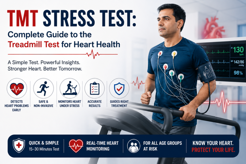
2D Echo vs ECG Difference: Which Test Do You Need?
Patients frequently ask about the 2D echo vs ECG difference — and understanding this distinction helps you appreciate why both tests are often complementary rather than interchangeable.
| Feature | ECG | 2D Echocardiography |
|---|---|---|
| What it measures | Electrical activity | Mechanical structure & function |
| What it shows | Rhythm, rate, ischemia signals | Chambers, valves, EF, wall motion |
| Technology used | Electrodes on skin | Ultrasound transducer |
| Procedure time | 5–10 minutes | 20–30 minutes |
| Radiation | None | None |
| Best for | Arrhythmias, heart attack diagnosis | Heart failure, valve disease, structural assessment |
| Cost | Lower | Moderate |
| Painful? | No | No |
The 2D echo vs ECG difference in practical terms: an ECG tells you how the heart is electrically conducting, while a 2D Echo tells you how the heart is physically moving and pumping. In most cardiac evaluations, both tests are performed together for a comprehensive assessment.
Echocardiography Procedure India: Availability and Access
The echocardiography procedure India has become increasingly accessible over the past decade. What was once available only in major metropolitan hospitals is now offered in well-equipped cardiac centres across smaller cities and towns.
The 2D echocardiography test is available across India at:
- Government medical college hospitals (often subsidised or free)
- District-level private hospitals and cardiac clinics
- Dedicated cardiology centres with trained sonographers
- Empanelled hospitals under PM-JAY and state health schemes
For BPL cardholders and patients enrolled under PM-JAY (Ayushman Bharat), SAST (Karnataka), or ESI schemes, the 2D echocardiography test is typically covered at zero out-of-pocket cost at empanelled cardiac hospitals. Patients visiting Hosapete for heart checkups can access 2D Echo at Jayam Hrudayalaya with expert cardiologist review.
The Heart Ultrasound Test: Safety and Suitability
The heart ultrasound test — which is exactly what a 2D Echo is — uses sound waves in the range of 1–5 MHz, far above the threshold of human hearing. These sound waves are entirely safe for human tissue.
There is no radiation involved. Unlike X-rays, CT scans, or nuclear stress tests, the 2D echocardiography test does not expose you to any ionising radiation whatsoever. This makes it completely safe for:
- Pregnant women (in fact, the same ultrasound technology is used for foetal imaging)
- Children and newborns (congenital heart disease evaluation)
- Elderly patients with multiple health conditions
- Patients requiring repeated monitoring over months or years
- Individuals with implanted devices such as pacemakers or ICDs
There are no known risks associated with diagnostic ultrasound at the frequencies used in echocardiography. It is one of the safest medical imaging modalities available.
When Does Your Cardiologist Recommend a 2D Echocardiography Test?
Your cardiologist will recommend a 2D echocardiography test in the following situations:
Symptomatic Indications:
- Breathlessness on exertion or at rest
- Chest pain or discomfort
- Heart palpitations or irregular heartbeat
- Swelling of feet, ankles, or abdomen (signs of heart failure)
- Fainting or near-fainting episodes
- Unexplained fatigue affecting daily activities
Following Other Abnormal Tests:
- Abnormal ECG findings suggesting ischemia, hypertrophy, or bundle branch block
- Chest X-ray showing enlarged heart shadow (cardiomegaly)
- Elevated BNP or NT-proBNP blood tests indicating cardiac strain
Monitoring Known Conditions:
- Serial assessment of known valve disease progression
- Post-heart attack evaluation of left ventricular function
- Monitoring of heart failure patients on treatment
- Follow-up after cardiac surgery or intervention
Preventive and Pre-Procedural:
- Pre-operative cardiac evaluation before major surgeries
- Baseline cardiac assessment in patients with diabetes, hypertension, or strong family history
- Evaluation before starting certain chemotherapy drugs that affect the heart
Why Expert Cardiologist Interpretation Matters
A 2D Echo report is only as valuable as the expertise of the cardiologist reading it. Echo interpretation requires years of training — recognising subtle wall motion abnormalities, grading valve lesions accurately, and distinguishing normal variants from pathological findings demands genuine clinical expertise.
At Jayam Hrudayalaya, every 2D echocardiography test is personally reviewed and reported by Dr. T. Sandeep (MBBS, MD, DNB Cardiology) — an interventional cardiologist with specialised training in echocardiographic interpretation and a clinical focus on comprehensive cardiac evaluation. This ensures that every finding, however subtle, is accurately identified and appropriately acted upon.
Conclusion
The 2D echocardiography test is the most comprehensive, safe, and informative non-invasive cardiac investigation available to patients today. It visualises your heart in motion, measures its pumping function, evaluates every valve, assesses wall thickness and motion, detects fluid collections, and identifies structural abnormalities — all in a single 20–30 minute session with no radiation, no pain, and no preparation required beyond arriving for your appointment.
If your doctor has recommended a 2D echocardiography test, do not delay. The information it provides is irreplaceable — and in many cases, it forms the cornerstone of your entire cardiac management plan. Your heart deserves to be seen clearly, assessed accurately, and treated expertly.