


Pacemaker
Implantation
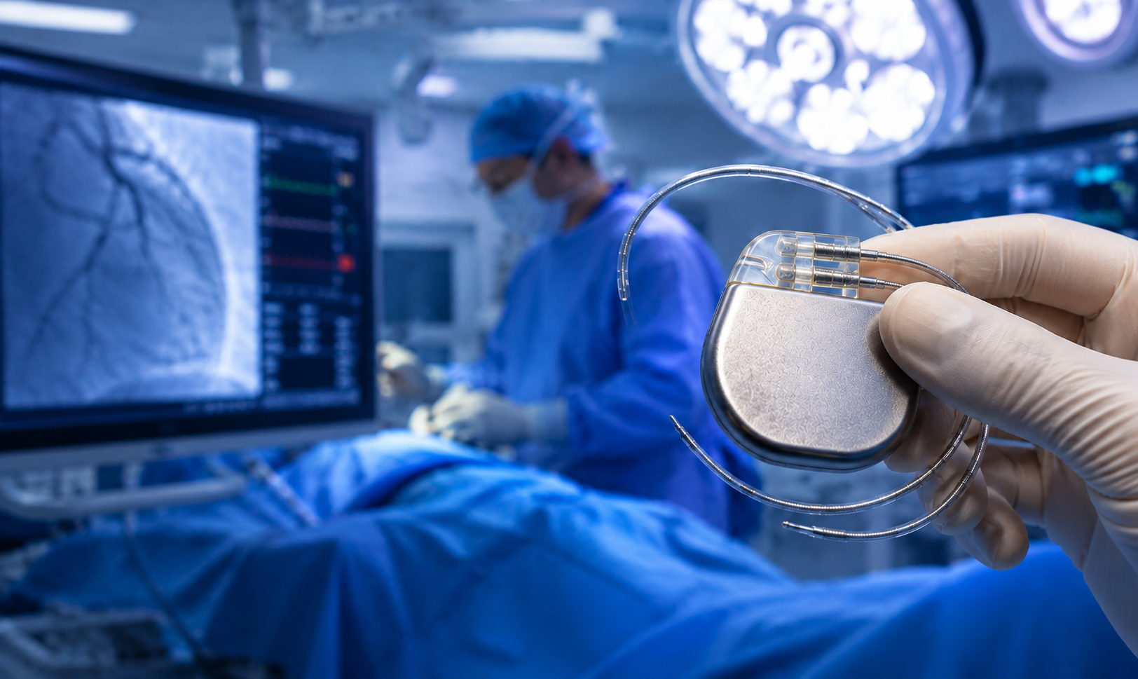
A small, life-saving electronic device surgically placed under the skin to regulate abnormal heart rhythms — giving your heart the precise electrical signal it needs to beat correctly.
72 BPM · VVI Mode · Battery 94%
Understanding the Device
What is a Pacemaker?
A pacemaker is a small battery-powered electronic device, roughly the size of a matchbox, implanted just beneath the skin of the upper chest. It continuously monitors the heart's electrical activity and delivers precisely timed electrical impulses through thin insulated wires called leads to stimulate the heart muscle when it beats too slowly, irregularly, or pauses altogether.
The normal human heart has its own natural pacemaker — the sinoatrial (SA) node — which generates electrical signals at 60–100 beats per minute. When this system malfunctions due to disease, ageing, or medication effects, an artificial pacemaker takes over this role reliably and safely.
At Jayam Hrudayalaya, pacemaker implantation is performed by our specialist cardiologist in a dedicated catheterisation laboratory using advanced fluoroscopic guidance, ensuring precise lead positioning and optimal device programming tailored to each patient's specific rhythm disorder.
Also Known As
Cardiac Pacemaker, Artificial Pacemaker, Implantable Pulse Generator (IPG), CRT-P (for cardiac resynchronisation therapy)
Performed By
Cardiac Electrophysiologist / Interventional Cardiologist in a sterile cath lab under fluoroscopic guidance
Anaesthesia Used
Local anaesthesia with conscious sedation; patient remains comfortable throughout — general anaesthesia is rarely required
Device Location
Generator placed in a pocket under the skin below the left or right collarbone; leads threaded through subclavian/cephalic vein into heart chambers
Battery Life
Modern pacemakers last 5–15 years depending on pacing dependency and settings; battery replacement requires a simple generator change procedure
Post-op Stay
Typically 24–48 hours for wound and rhythm monitoring; device is programmed and tested before discharge with a detailed follow-up plan
Step-by-Step Procedure
How Pacemaker Implantation is Performed
Pre-procedure Preparation
The patient is positioned on the cath lab table and connected to continuous ECG monitoring. The chest area (usually left side) is cleaned, shaved, and draped in sterile fashion. An IV line is placed and sedation is given to keep the patient comfortable and relaxed throughout the procedure.
Local Anaesthesia & Skin Incision
Local anaesthetic (lignocaine) is injected beneath the collarbone to numb the area completely. A 4–6 cm incision is made just below the clavicle. A pocket is carefully created in the subcutaneous tissue to accommodate the pacemaker generator without causing discomfort after healing.
Venous Access & Lead Introduction
The subclavian or cephalic vein is accessed to introduce the pacemaker lead(s). A guidewire and introducer sheath are used to create a smooth pathway into the vein. One or two leads are then carefully advanced through the venous system toward the heart under continuous X-ray fluoroscopy.
Lead Positioning in the Heart
The lead tip is positioned precisely inside the target heart chamber — the right ventricle (for single-chamber), or both right atrium and right ventricle (for dual-chamber pacemakers). The lead is actively or passively fixed using a helix tip or tines. Fluoroscopic images confirm correct placement.
Lead Testing & Threshold Measurement
Each lead is connected to an external pacing system analyser (PSA). Electrical measurements are taken: sensing threshold, pacing threshold, and lead impedance. These values confirm optimal lead position before final fixation and generator connection.
Generator Connection & Pocket Closure
The tested leads are connected to the pacemaker generator, which is placed securely into the pre-formed subcutaneous pocket, sutured in place, and the incision closed in layers using absorbable sutures covered with a sterile dressing. Total procedure time: 1–2 hours.
Device Programming & Verification
After closure, the pacemaker is programmed wirelessly. Parameters including base rate, pacing mode, sensitivity, and output are set to the patient's specific requirements. A chest X-ray confirms lead positions and rules out pneumothorax before the patient is shifted to the recovery ward.
Device Variants
Types of Pacemakers
Single-Chamber Pacemaker
One lead placed in either the right ventricle or right atrium. Simple, cost-effective, and suitable for most bradycardia cases.
- VVI or AAI pacing mode
- One lead — simpler implant procedure
- Suitable for permanent AF + slow rate
- Smaller generator — ideal for elderly
- Battery life: 8–12 years
Dual-Chamber Pacemaker
Two leads — one in the right atrium and one in the right ventricle — coordinate timing between upper and lower chambers, mimicking natural conduction.
- DDD pacing mode — most physiological
- Maintains atrio-ventricular (AV) synchrony
- Prevents "pacemaker syndrome"
- Preferred in AV block and sick sinus syndrome
- Battery life: 7–10 years
Biventricular Pacemaker (CRT-P)
Cardiac Resynchronisation Therapy — three leads coordinate both ventricles and the right atrium. Used in heart failure with bundle branch block.
- Improves ejection fraction in selected patients
- Reduces heart failure hospitalisations
- LV lead placed via coronary sinus
- Can be combined with ICD (CRT-D)
- Battery life: 5–8 years
Leadless Pacemaker
A miniaturised self-contained device delivered directly into the right ventricle via catheter — completely eliminating leads and subcutaneous pocket.
- No chest incision or pocket required
- Eliminates lead-related complications
- Ideal for infection-risk patients
- Micra™ AV — dual-chamber sensing
- Battery life: 10–14 years
ICD (Implantable Cardioverter-Defibrillator)
Combines pacemaker function with the ability to deliver a high-energy shock to terminate life-threatening ventricular arrhythmias. For patients at high risk of sudden cardiac death.
- Monitors for VF/VT continuously
- Delivers ATP (anti-tachycardia pacing) first
- Delivers DC shock if ATP fails
- Indicated for EF ≤35% with symptoms
- Battery life: 5–9 years
Temporary Pacemaker
External non-implanted pacing used in acute settings — complete heart block after MI, drug toxicity, or as a bridge to permanent device in unstable patients.
- Transvenous or transcutaneous pacing
- Used in ICU / emergency setting
- Lead placed via femoral or jugular vein
- Duration: hours to days
- Bridge to permanent device or recovery
Device Anatomy
Components of a Pacemaker System
Pulse Generator (IPG)
The main body containing the battery (lithium-iodide) and electronic circuitry. It generates electrical impulses, processes sensed signals, and stores diagnostic data. Hermetically sealed in a titanium casing — safe for most environments.
Pacing Leads
Thin, insulated wires connecting the generator to the heart muscle. Each lead has an electrode at the tip that senses intrinsic activity and delivers stimuli. Available in active-fixation (helix) or passive-fixation (tined) designs.
Programmer (External)
A bedside device communicating wirelessly with the implanted pacemaker via a wand placed over the device pocket. Allows cardiologists to adjust pacing rates, sensitivity, output energy, and modes non-invasively at each follow-up.
Remote Monitoring System
Modern pacemakers include wireless telemetry allowing nightly data transmission to a secure server. Cardiologists can review arrhythmia logs, battery status, and lead integrity remotely — reducing clinic visits while increasing safety.
Rate Response Sensor
An accelerometer inside the generator that detects physical activity and automatically increases the pacing rate to meet the body's increased oxygen demand during exercise — allowing patients to remain active with good quality of life.
MRI-Conditional Design
Modern pacemakers are engineered to be "MRI-conditional" — they can safely undergo MRI scans under specific conditions. Today's generation allows patients to receive important diagnostic imaging without compromising device safety.
Medical Conditions Treated
When is a Pacemaker Needed?
Sinus Bradycardia
Persistently slow heart rate (<40 bpm) causing dizziness, fatigue, or fainting that does not respond to medication adjustment.
Sick Sinus Syndrome
SA node dysfunction causing alternating slow and fast rhythms, sinus pauses, or sinoatrial exit block with significant symptoms.
Complete Heart Block
Total electrical disconnection between atria and ventricles — ventricles beat independently at a dangerously slow rate of 20–40 bpm.
High-Degree AV Block
2nd degree Mobitz Type II AV block — predictable dropped beats that can progress to complete heart block; pacemaker is mandatory.
Recurrent Syncope
Frequent fainting episodes with documented prolonged asystole (>3 seconds) or severe bradycardia during tilt testing or event monitors.
Drug-Induced Bradycardia
When essential cardiac medications cause symptomatic bradycardia that cannot be discontinued or dose-reduced safely.
Heart Failure with LBBB
Reduced ejection fraction (≤35%) with left bundle branch block (QRS ≥130 ms) — biventricular pacing resynchronises ventricular contraction.
Post-Cardiac Surgery Bradycardia
Persistent AV block or sinus node dysfunction following cardiac surgery that has not resolved within 5–7 days of the procedure.
Pacing Modes Reference
Pacemaker Modes — NBG Code Explained
| Mode Code | Chamber Paced | Chamber Sensed | Response | Best Indicated For | Notes |
|---|---|---|---|---|---|
| VVI | Ventricle (R) | Ventricle (R) | Inhibited | Permanent AF + slow ventricular rate | Single Lead |
| AAI | Atrium (R) | Atrium (R) | Inhibited | Sinus node dysfunction with intact AV conduction | Single Lead |
| DDD | Both (A + V) | Both (A + V) | Inhibit / Trigger | AV block, sick sinus syndrome — most physiological | Gold Standard |
| DDDR | Both (A + V) | Both (A + V) | Inhibit / Trigger + Rate Response | Active patients needing rate adaptation during exercise | Rate Adaptive |
| VOO / AOO / DOO | V / A / Both | None | None (Asynchronous) | Temporary pacing; near strong electromagnetic interference | Magnet Mode |
| CRT (BiV DDD) | Both Ventricles + RA | Both (A + V) | Inhibit / Trigger | Heart failure with LBBB, EF ≤35% | 3 Leads |
Clinical Considerations
Benefits & Risks of Pacemaker Implantation
Benefits of the Procedure
Why pacemaker implantation transforms quality of life
- ✓Complete elimination of fainting, dizziness, and extreme fatigue caused by bradycardia within days of implant
- ✓Life-saving in complete heart block — prevents sudden cardiac arrest due to asystole or very slow escape rhythm
- ✓Enables safe use of essential medications that were previously dose-limited by bradycardia
- ✓Dramatic improvement in exercise capacity and quality of life — patients return to full normal activity
- ✓CRT pacemakers improve heart failure symptoms, reduce hospitalisations, and can improve ejection fraction
- ✓Modern devices are MRI-compatible, remote-monitored, and last 8–15 years with minimal maintenance
- ✓Minimally invasive — no open-chest surgery, short hospital stay, and rapid recovery
Possible Risks & Complications
Uncommon but important risks discussed during informed consent
- !Pocket haematoma — blood collection at implant site; most resolve spontaneously, rarely requires drainage (<2%)
- !Pneumothorax — accidental lung puncture during subclavian vein access; managed with chest drain if large (<1%)
- !Lead dislodgement — lead tip moves from initial position; detected on post-op X-ray; requires repositioning (<2%)
- !Device infection — skin or pocket infection; rare but serious; may require device explantation (<1%)
- !Pacemaker syndrome — in VVI pacing, loss of AV synchrony causing fatigue and hypotension; upgraded to dual-chamber
- !Cardiac perforation — rare lead tip perforation of ventricular wall (<0.5%); managed immediately in cath lab
- !Electromagnetic interference — strong external EM sources may temporarily affect device function
Before the Procedure
How to Prepare for Pacemaker Implantation
Fasting Guidelines
Fast for 4–6 hours before the procedure. A sip of water is permitted for essential medications. Emergency implants proceed without fasting delay when the patient is haemodynamically unstable.
Medications to Review
Inform the cardiologist about all medications — anticoagulants (warfarin stopped 3–5 days prior; novel anticoagulants paused 24–48 hrs), aspirin, insulin, and antibiotics. Never stop medications without explicit medical advice.
Blood Tests Required
Full blood count, renal function, coagulation profile (PT-INR), blood group and cross-match, blood sugar, and ECG. Chest X-ray and echocardiogram are reviewed to plan device selection and lead positioning.
Skin Preparation
The chest and shoulder area will be cleaned and shaved in the procedure room. Avoid applying creams, lotions, or deodorant on the day of the procedure to reduce infection risk at the implant site.
IV Access & Antibiotics
Prophylactic IV antibiotics (typically cefazolin) are given 30–60 minutes before skin incision to prevent pocket infection — one of the most important pre-procedure steps in pacemaker surgery.
Informed Consent & Family
The cardiologist will explain the device type, procedure steps, potential risks, and post-implant care. Family members are counselled on emergency contact numbers and warning signs requiring immediate medical attention.
Post-Implant Care
Recovery After Pacemaker Implantation
In Hospital — First 24–48 Hours
You will be monitored in the cardiac ward with continuous ECG. A chest X-ray confirms lead positions and rules out complications. The pacemaker is checked with the programmer before discharge. Most patients feel immediate relief of bradycardia symptoms.
Wound Care at Home
Keep the incision clean and dry for 5–7 days. Avoid soaking the area in water (no baths, swimming) until fully healed. Watch for signs of infection: redness, swelling, warmth, discharge, or fever. Attend wound review at 7–10 days for suture removal.
Arm & Activity Restrictions
Avoid lifting the implant-side arm above shoulder level for 4–6 weeks to allow leads to heal firmly in place. No heavy lifting (>2–3 kg) for 4–6 weeks. Gentle walking from day 1. Most patients return to desk work within 1 week.
Device ID Card & Documentation
You will receive a pacemaker identification card with your device model, serial number, settings, and cardiologist details. Carry this card at all times and present it before any medical procedure, security screening, or MRI scan.
Follow-up Visits
Follow-up device checks at 4–6 weeks, 3 months, 6 months, then annually. Each visit includes ECG, device interrogation, battery status check, and reprogramming if needed. Remote monitoring transmissions are reviewed between visits.
Battery Replacement (Generator Change)
When the battery reaches end-of-life (typically 8–12 years), a generator change is performed under local anaesthesia. The old generator is removed, leads are tested and reused if functioning well, and a new generator connected and programmed within 24 hours hospital stay.
Long-term Guidance
Living with Your Pacemaker
Mobile Phones & Electronics
Modern pacemakers are well-shielded. Use your mobile phone on the opposite side to the pacemaker. Keep the phone at least 15 cm from the device. Wireless earphones, laptops, and household appliances are generally safe.
Exercise & Physical Activity
Most patients can exercise freely. Avoid contact sports or activities with high risk of direct chest impact. Swimming, cycling, walking, and yoga are safe. The rate-response feature adjusts pacing rate during exercise automatically.
Air Travel & Security
Pacemakers are safe for air travel. Inform airport security and present your ID card; request a hand-wand search if unsure. The device will not be damaged by airport security equipment, but metal detectors may trigger an alarm.
Equipment & Machinery
Avoid arc welding and strong industrial magnets. Household appliances including microwaves, induction cookers, and power tools are safe to use. Inform your doctor before any electrosurgical (diathermy) procedure.
MRI & Medical Procedures
If your device is MRI-conditional (most modern pacemakers), MRI scans can be safely performed under specific conditions with device reprogramming. Always inform the radiologist and MRI technician of your pacemaker before any scan.
Diet & Lifestyle
A heart-healthy lifestyle reduces the risk of further cardiac disease. Eat a balanced diet low in saturated fat and salt. Quit smoking, maintain a healthy weight, and manage diabetes and hypertension diligently.
Frequently Asked Questions
Pacemaker implantation is considered a minor surgical procedure. It is performed under local anaesthesia with mild sedation — the patient is awake and comfortable throughout. There is no need for general anaesthesia or opening of the chest. The procedure takes 1–2 hours, and most patients are discharged within 24–48 hours, resuming normal daily activities within 1–2 weeks.
Most patients do not feel individual pacemaker stimulations. In the weeks after implantation, some notice a mild awareness of the device under the skin — the generator creates a small visible or palpable bulge under the collarbone, especially in thin individuals. This is entirely normal. Rarely, patients feel a mild twitching sensation which can be resolved by reprogramming at a follow-up visit.
Modern pacemaker batteries typically last 8–12 years, depending on how often the device is required to pace. At every follow-up, the programmer reads the remaining battery life accurately. When the battery reaches "elective replacement indicator" (ERI) status — usually 3–6 months before end-of-life — a generator change is planned as an elective procedure. The leads are usually reused and the new generator is connected and programmed in a short, simple procedure.
Yes — microwave ovens, induction cookers, electric blankets, and most standard household appliances are safe with modern pacemakers. These devices operate at frequencies and field strengths that do not interfere with contemporary pacemakers. The main equipment to avoid includes industrial arc welding equipment, large permanent or electro-magnets, and certain medical equipment such as electrosurgical diathermy units. Always carry your device ID card and inform medical professionals before any procedure.
It depends on your underlying condition. Patients with complete heart block or very slow intrinsic rhythms are highly dependent on the pacemaker — their heart relies on the device for every beat. Other patients may only need occasional pacing support, with their own natural rhythm taking over most of the time. Your cardiologist will monitor your intrinsic rhythm at follow-up visits. Dependency is not a concern — the pacemaker is designed to function continuously and reliably for many years.
Yes — at Jayam Hrudayalaya, pacemaker implantation is available under government BPL (Below Poverty Line) assistance schemes, making this life-saving treatment accessible to all patients. We also accept most major insurance providers and government health schemes. Our administrative team assists with pre-authorisation, documentation, and claim processing. Please visit the BPL Scheme page on our website for eligibility details and required documents.
Contact emergency immediately if you experience: return of fainting, severe dizziness, or extreme fatigue (may indicate lead dislodgement or device malfunction); chest pain, shortness of breath, or palpitations; fever above 38°C with redness, swelling, or discharge at the wound site (signs of infection); visible swelling or a pulsating lump at the wound site; persistent rhythmic hiccups (may indicate lead perforation). Always carry your pacemaker ID card so emergency teams can identify your device immediately.