


TMT — Treadmill Test Exercise Stress Electrocardiography
A dynamic cardiac stress test that monitors your heart's electrical activity, blood pressure, and symptoms while you exercise — uncovering hidden heart disease invisible at rest.
The Treadmill Test (TMT), also known as the Exercise Stress Test or Stress ECG, is a diagnostic cardiac investigation that assesses how your heart performs under physical stress. It records your ECG, heart rate, and blood pressure continuously as you walk or jog on a motorised treadmill at progressively increasing speeds and inclines.
At rest, coronary artery disease may not be evident on a standard ECG. However, when the heart is made to work harder through exercise, areas of reduced blood supply (ischaemia) become apparent through characteristic changes in the ECG tracing — particularly ST-segment depression or elevation.
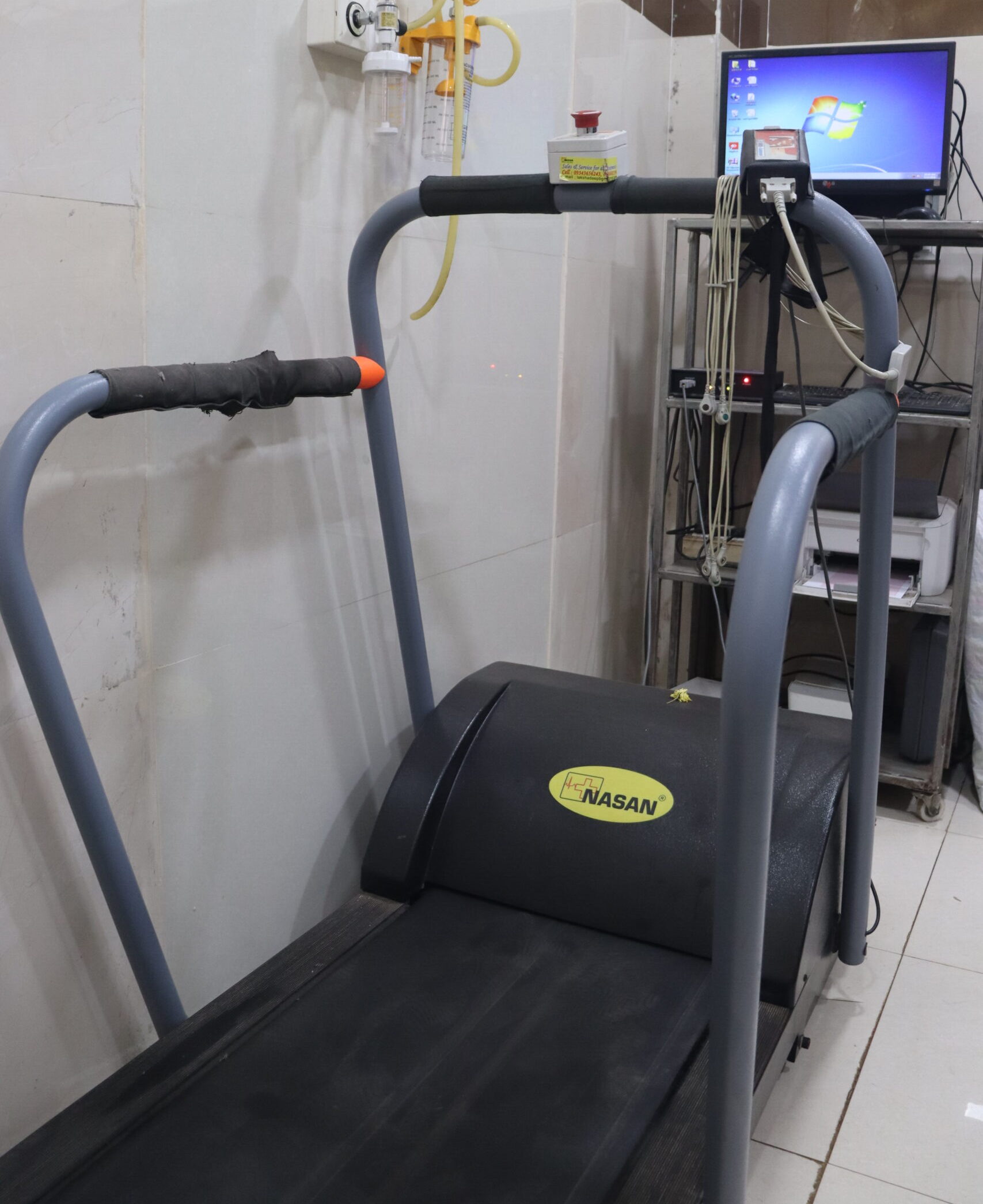
At Jayam Hrudayalaya, our TMT laboratory is equipped with digital 12-lead ECG systems, automated blood pressure monitoring, emergency resuscitation equipment, and trained cardiac technicians with a cardiologist present throughout the test for maximum safety.
Detects Hidden Ischaemia
Reveals coronary artery disease that remains invisible on resting ECG or 2D Echo
Dynamic Monitoring
Continuous 12-lead ECG + blood pressure every 2–3 minutes throughout the test
Safe & Supervised
Cardiologist present throughout; immediate termination criteria defined
Functional Assessment
Measures exercise capacity in METs — a key predictor of cardiac prognosis
Also Called
Exercise Stress Test, Stress ECG, Exercise Tolerance Test, EST
Duration
15–30 minutes total including prep, exercise, and recovery monitoring
Target HR
85% of Maximum Predicted Heart Rate = 220 minus patient's age
Protocol Used
Standard Bruce Protocol — 7 stages, each 3 minutes duration
What's Monitored
12-lead ECG, Heart Rate, Blood Pressure, Symptoms, SpO2
Who's Present
Cardiac technician + Cardiologist present for full duration of test
The standard Bruce Protocol consists of 7 stages, each lasting 3 minutes, with increasing treadmill speed and incline. The test is stopped when the target heart rate is reached, symptoms develop, or ECG changes occur.
| Stage | Speed (mph) | Speed (km/h) | Grade (%) | Duration | MET Level | Intensity |
|---|---|---|---|---|---|---|
| Stage 1 | 1.7 | 2.7 | 10% | 3 min | ~4–5 | Very Light |
| Stage 2 | 2.5 | 4.0 | 12% | 3 min | ~7 | Light |
| Stage 3 | 3.4 | 5.4 | 14% | 3 min | ~10 | Moderate |
| Stage 4 | 4.2 | 6.7 | 16% | 3 min | ~13 | Moderate-Hard |
| Stage 5 | 5.0 | 8.0 | 18% | 3 min | ~16 | Hard |
| Stage 6 | 5.5 | 8.9 | 20% | 3 min | ~19 | Very Hard |
| Stage 7 | 6.0 | 9.6 | 22% | 3 min | ~22 | Maximal |
MET Load Progression by Stage
Here is exactly what happens during your TMT appointment at Jayam Hrudayalaya:
Registration & Clinical Review
You will be registered, your medical history reviewed, and the cardiologist will assess your resting ECG and BP. Any medications, symptoms, or risk factors are noted. The procedure is explained and informed consent obtained.
Electrode Placement & Skin Preparation
The technician will clean the skin with alcohol and lightly abrade electrode sites for good contact. 10 electrodes are attached to your chest, shoulders, and legs to record a full 12-lead ECG. Blood pressure cuff is applied on the left arm.
Resting Baseline Recording
A complete resting 12-lead ECG and blood pressure are recorded before exercise begins. This forms the baseline against which exercise changes are compared. Symptoms at rest are noted.
Exercise Phase — Bruce Protocol Stages
The treadmill starts at Stage 1 (1.7 mph, 10% grade) and automatically advances every 3 minutes. ECG is monitored continuously; blood pressure is recorded every 3 minutes. You report any symptoms (chest pain, breathlessness, dizziness, palpitations) immediately. The goal is to reach 85% of your maximum predicted heart rate (220 – age).
Recovery Phase Monitoring
After exercise stops, you rest on the couch or continue slow walking. ECG and BP are monitored continuously for 8 minutes. Many significant changes (ST changes, arrhythmias) appear only in the recovery phase — making this period equally important as exercise itself.
Cardiologist Interpretation
The cardiologist analyses the ECG tracings from all phases — baseline, each exercise stage, and recovery. Key findings including ST changes, arrhythmias, BP response, heart rate recovery, and exercise capacity in METs are assessed and documented.
Report & Consultation
A detailed TMT report is provided with a conclusion of Positive, Negative, or Inconclusive. The cardiologist discusses findings, implications, and the next steps — which may include medical management, further imaging (coronary angiogram, stress echo), or lifestyle advice.
Your cardiologist may recommend a Treadmill Test in these clinical situations:
Exertional Chest Pain
Chest tightness or pain that occurs specifically during physical activity or exercise
Suspected CAD
Evaluation of suspected coronary artery disease in patients with risk factors
Exertional Palpitations
Heart rhythm abnormalities triggered by exercise — to identify exercise-induced arrhythmias
Unexplained Breathlessness
Dyspnoea on exertion not explained by respiratory causes — cardiac origin evaluation
Post-MI Assessment
Functional assessment 4–6 weeks after a heart attack to guide cardiac rehabilitation
Known CAD Follow-up
Monitoring response to medical therapy or assessing residual ischaemia post-procedure
Pre-sport Clearance
Cardiac fitness assessment before starting a vigorous exercise programme or competitive sports
Pre-operative Evaluation
Risk stratification before major non-cardiac surgical procedures in high-risk patients
Drug Therapy Monitoring
Assessing effectiveness of anti-anginal medications and rate-control drugs during exercise
Exertional Syncope
Blackouts or near-fainting episodes occurring during or immediately after physical exertion
TMT must not be performed in certain high-risk conditions. Your cardiologist will screen you carefully before the test:
🚫 Absolute Contraindications
Test must NOT be performed in these conditions
⚠️ Relative Contraindications
Test may proceed with extra caution & cardiologist decision
Understanding your TMT report — what the cardiologist looks for at each stage and in recovery:
| Parameter | Normal Response | Abnormal / Positive Finding | Clinical Significance |
|---|---|---|---|
| ST Segment | Flat or upsloping, no change >1mm | ≥1mm horizontal/downsloping ST depression | Most reliable indicator of myocardial ischaemia due to coronary artery disease |
| ST Elevation | Not present in exercise leads | ≥1mm ST elevation in non-Q leads | Severe ischaemia, vasospasm, or extensive wall motion abnormality — urgent evaluation needed |
| Heart Rate Response | Progressive rise; 85% MPHR achieved | Chronotropic incompetence (<80% MPHR) | Inability to raise HR appropriately — indicates sinus node dysfunction or severe LV dysfunction |
| Blood Pressure Response | Systolic rises 10–50 mmHg per stage | Drop >10 mmHg = hypotensive response Rise >250/115 = hypertensive | BP drop during exercise is an ominous sign indicating severe LV dysfunction or significant CAD |
| Exercise Capacity (METs) | >10 METs = good functional capacity | <5 METs = poor prognosis 5–7 METs = intermediate risk | One of the strongest independent predictors of cardiovascular mortality and overall prognosis |
| Heart Rate Recovery (HRR) | HR drops ≥12 bpm at 1 min after exercise | HRR <12 bpm = abnormal autonomic response | Impaired HRR is associated with increased mortality independent of ST changes |
| Duke Treadmill Score | Score ≥5 = Low risk | Score ≤-11 = High risk (annual mortality >5%) -10 to +4 = Intermediate | DTS = Exercise time – (5 × ST deviation) – (4 × angina index). Most validated TMT risk score |
| Symptoms During Test | None, or mild breathlessness | Anginal chest pain replicating symptoms | Symptom-limited test with typical angina is highly suggestive of significant CAD even without ST changes |
| Arrhythmias | Occasional isolated ectopics only | Ventricular tachycardia / frequent PVCs / AF | Exercise-induced VT or SVT warrants further evaluation with EP study or advanced imaging |
A TMT is reported as POSITIVE (indicating probable significant coronary artery disease) when any of these criteria are met:
ST Depression ≥1 mm
Horizontal or downsloping ST depression of ≥1 mm (0.1 mV) measured 60–80 ms after the J-point in ≥2 contiguous leads. This is the most classic positive criterion for myocardial ischaemia.
ST Elevation ≥1 mm
ST elevation ≥1 mm in leads without prior Q waves (excluding aVR and V1). This indicates transmural ischaemia or vasospasm and is a high-risk finding requiring immediate evaluation.
Typical Anginal Chest Pain
Development of typical anginal chest pain (central, heavy, radiating) during the test — especially if it requires test termination. Anginal symptoms significantly increase the predictive value of any ST changes.
Significant BP Drop
A fall in systolic blood pressure of >10 mmHg below baseline during progressive exercise stages — suggesting severe LV dysfunction, left main disease, or three-vessel coronary artery disease.
Exercise-Induced VT
Sustained or non-sustained ventricular tachycardia (≥3 consecutive beats) triggered by exercise. This is a serious finding requiring urgent further investigation including electrophysiology study.
Low Exercise Capacity
Inability to complete Stage 2 of the Bruce Protocol (<7 METs) combined with ST changes indicates high-risk coronary artery disease with significant impact on quality of life and prognosis.
Proper preparation ensures accurate results and maximum safety. Follow these instructions carefully before your TMT appointment:
Fast for 3–4 Hours
Do not eat a heavy meal within 3–4 hours before the test. A light snack 1–2 hours prior is acceptable. Avoid caffeine (tea, coffee, energy drinks) for at least 4 hours as it affects heart rate response.
Medication Instructions
This is critical — ask your cardiologist specifically about beta-blockers and rate-limiting calcium channel blockers. They may need to be withheld for 24–48 hours before the test as they blunt the heart rate response and can cause false-negative results.
Comfortable Footwear
Wear proper athletic or sports shoes with non-slip soles. Do not wear slippers, sandals, or formal shoes. Loose, comfortable sportswear or tracksuit is recommended for the test.
Avoid Smoking
Do not smoke for at least 3 hours before the test. Nicotine affects heart rate and vascular tone, which can interfere with the accuracy of the exercise ECG response.
Inform About Symptoms
Inform the cardiologist if you have had recent chest pain, breathlessness, or palpitations at rest in the past 48 hours — the test may need to be deferred for your safety.
Carry Previous Records
Bring your previous ECG reports, 2D Echo reports, angiogram reports, and medication list. This helps the cardiologist compare changes and interpret the TMT findings in clinical context.
Choosing the right cardiac stress test depends on your clinical situation, risk profile, and pre-existing ECG findings:
🏃 TMT (Treadmill Test)
Exercise Stress ECG
📡 Stress Echocardiogram
Exercise / Dobutamine Echo
🔬 Coronary Angiogram (CAG)
Gold Standard — Invasive
Book Your TMT at Jayam Hrudayalaya
Expert cardiac stress testing with cardiologist supervision, immediate results, and BPL scheme benefits for eligible patients.