


Coronary
Angiogram
(Angiography)
The gold standard X-ray procedure that maps your coronary arteries with precision — revealing blockages before they become life-threatening. Led by Dr. T. Sandeep, DNB (Cardiology).
Dr. T. Sandeep
Interventional CardiologistWhat is a Coronary Angiogram?
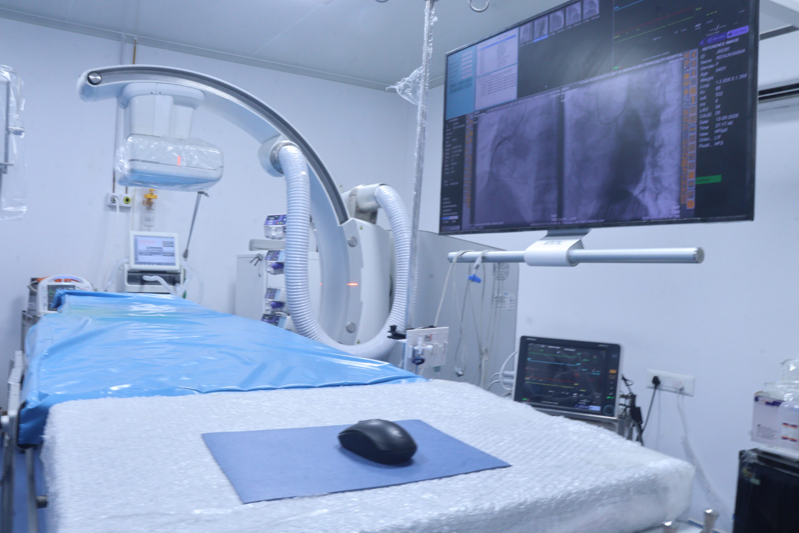
A cathlab-based X-ray imaging procedure that gives your cardiologist the most accurate, real-time view inside your coronary arteries.
A Coronary Angiogram (also called Coronary Angiography or Cardiac Catheterisation) is a minimally invasive diagnostic procedure performed in a Catheterisation Laboratory (Cathlab). A thin, flexible tube called a catheter is guided through the radial artery (wrist) or femoral artery (groin) to the opening of your coronary arteries.
A contrast dye (iodine-based) is then injected through the catheter, making the coronary arteries instantly visible on real-time X-ray screens (fluoroscopy). This allows Dr. T. Sandeep to identify blockages, narrowing (stenosis), and lesions with pinpoint accuracy — revealing exactly where, how many, and how severe the blockages are.
The angiogram is the definitive guide for treatment decisions — determining whether a patient requires only medication, angioplasty (PTCA/stenting), or bypass surgery (CABG).
- Minimally invasive — no surgical incision, only a small catheter insertion
- Gold standard accuracy — detects blockages as small as 1–2 mm in diameter
- Immediate results — findings reviewed live on screen with instant decision
- Procedure lasts 15–30 minutes under local anaesthesia (you stay awake)
- Same-day discharge possible with radial (wrist) approach
- Free of cost for eligible BPL cardholders under PM-JAY, SAST & other schemes
Did you know? A coronary angiogram can detect blockages as small as 1–2 mm — far beyond what ECG, 2D Echo, or TMT can reveal. It remains the definitive diagnostic tool for coronary artery disease worldwide.
When Is a Coronary
Angiogram Recommended?
Your cardiologist may advise a coronary angiogram when symptoms, test results, or clinical risk suggest significant coronary artery disease.
Chest Pain (Angina)
Persistent or worsening chest pain, pressure, or tightness — especially on exertion — that suggests coronary artery blockage requiring direct visualisation and assessment.
Abnormal Stress Test (TMT)
If your Treadmill Test shows significant ST changes, an angiogram confirms the exact location and severity of coronary disease and guides further treatment decisions.
Heart Attack (STEMI / NSTEMI)
Emergency angiogram is performed immediately after a heart attack to identify the culprit blocked artery and guide emergency angioplasty (primary PTCA) to restore blood flow.
Reduced Ejection Fraction
When 2D Echo shows a weak heart muscle (low EF), an angiogram determines whether blocked arteries are the ischaemic cause and whether revascularisation can restore function.
Pre-Surgical Cardiac Evaluation
Before major cardiac or non-cardiac surgery in patients with risk factors, an angiogram assesses coronary artery status to ensure safe surgical planning and management.
Unexplained Heart Failure
When the cause of heart failure is uncertain, an angiogram rules out or confirms coronary artery disease as the underlying ischaemic aetiology requiring intervention.
Types of Coronary Angiogram
The procedure can be performed via different access routes. At Jayam Hrudayalaya, we prefer the modern radial (wrist) approach for faster recovery and minimal discomfort.
Radial Approach (Wrist)
The catheter is inserted through the radial artery at the wrist. This is the modern gold-standard technique with the lowest complication rate.
- No bed rest required after procedure
- Walk immediately after the procedure
- Same-day discharge in most patients
- Significantly lower bleeding risk
- Greater patient comfort and satisfaction
Femoral Approach (Groin)
The catheter is inserted through the femoral artery in the groin. Used when radial access is not anatomically feasible or in specific clinical situations.
- Requires 4–6 hours bed rest post-procedure
- Leg must remain straight during recovery
- Overnight stay usually recommended
- Slightly higher access site bleeding risk
- Larger catheter sizes possible if needed
Diagnostic Angiogram
A purely diagnostic procedure to map all coronary arteries and identify the presence, location, and severity of blockages. The findings guide all subsequent treatment decisions.
- No intervention performed in same sitting
- Used when diagnosis needs confirmation
- Results discussed with family after procedure
- Treatment planned based on findings
Ad-Hoc Angioplasty
When a significant blockage is identified during angiogram, the cardiologist may proceed directly to angioplasty (PTCA/stenting) in the same session — called ad-hoc angioplasty.
- Avoids a second separate procedure
- Reduces overall treatment delay significantly
- One anaesthesia episode and recovery
- Preferred in stable suitable anatomy cases
How the Procedure
Is Performed
From preparation to discharge — exactly what happens during your coronary angiogram at Jayam Hrudayalaya's state-of-the-art Cathlab.
Pre-Procedure Preparation
You will be asked to fast for 4–6 hours before the procedure. Blood tests (CBC, renal function, coagulation profile), ECG, and prior reports are reviewed. An IV line is placed for medications. The access site is cleaned and a mild sedative is administered to help you feel relaxed and calm.
⏱ 30–45 minutes preparationLocal Anaesthesia & Arterial Access
Local anaesthetic (lignocaine) is injected at the insertion site to completely numb the area — you will feel only mild pressure, not pain. A small puncture is made and a short plastic sheath tube is inserted into the radial or femoral artery. The catheter is threaded through the sheath and carefully advanced toward the heart under continuous real-time X-ray (fluoroscopy) guidance.
⏱ 5–10 minutesContrast Dye Injection & X-Ray Imaging
The catheter tip is precisely positioned at the opening of each coronary artery. A small amount of contrast dye is injected — you may feel a brief warm or flushing sensation for a few seconds, which is completely normal. Multiple high-resolution X-ray images are captured from different angles, creating a complete map of all your coronary arteries, precisely revealing any blockages, stenosis, or lesions.
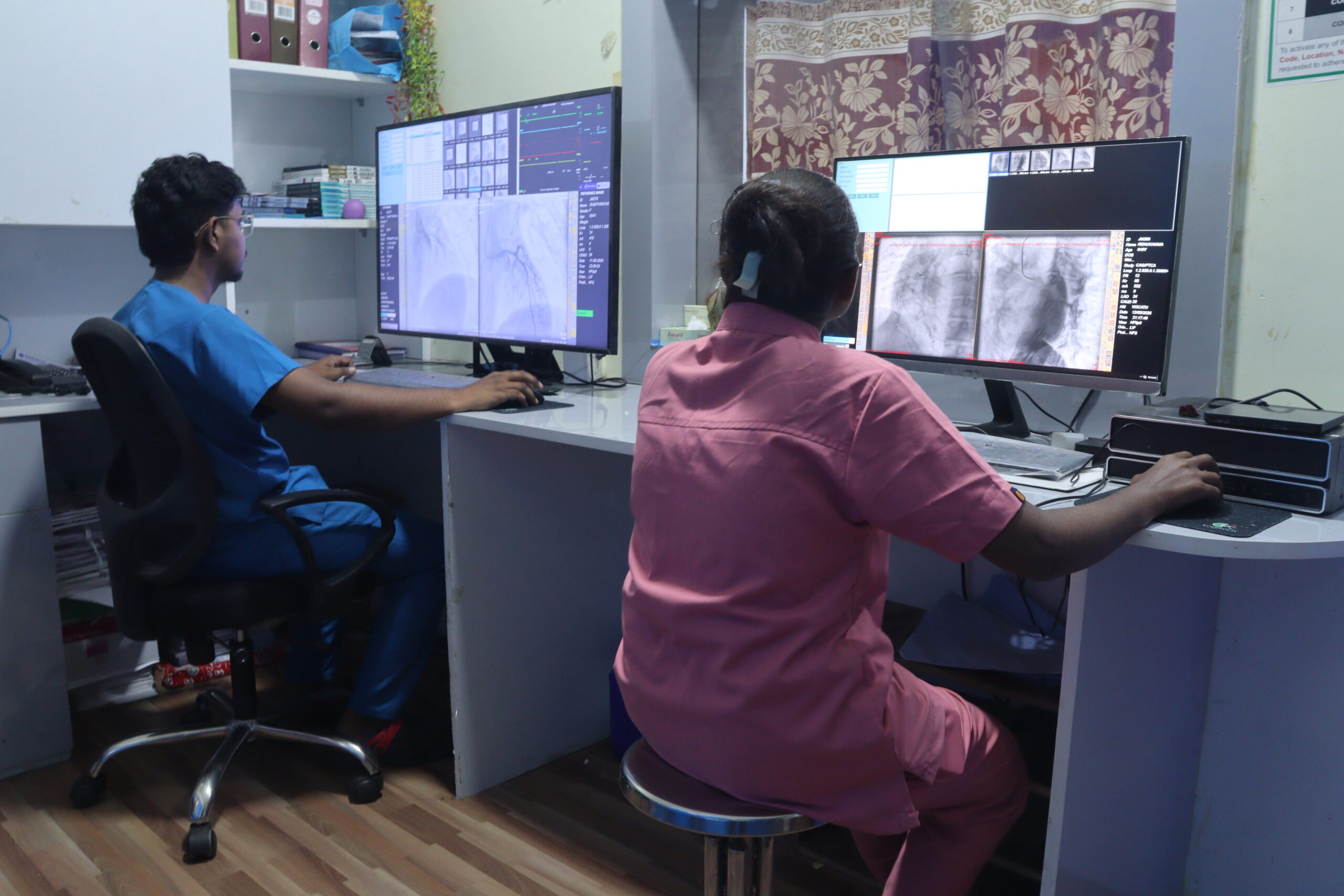
⏱ 15–30 minutes imagingReal-Time Assessment & Treatment Decision
Dr. T. Sandeep reviews the angiogram images live on high-resolution screens in the Cathlab. If a significant blockage is found (typically >70% narrowing), the decision is made immediately — whether to proceed with angioplasty in the same session (ad-hoc), plan a separate procedure, or refer for bypass surgery (CABG). The findings and options are explained clearly to you and your family right after.
⏱ 5–10 minutes assessmentCatheter Removal & Access Site Closure
After imaging is complete, the catheter and sheath are carefully removed. For radial (wrist) access, a TR (Trans-Radial) compression band is applied to the wrist for 2–4 hours. For femoral (groin) access, manual pressure is applied for 10–15 minutes or a vascular closure device is used. You are then transferred to the cardiac recovery area.
⏱ 5–10 minutesRecovery Monitoring & Discharge
Vital signs are continuously monitored for 2–4 hours in the cardiac recovery unit. The wrist compression band is gradually deflated and removed. If no angioplasty was performed, most radial-approach patients are discharged the same day with detailed discharge instructions, prescribed medications, and a follow-up appointment. Femoral approach requires overnight observation.
⏱ 2–6 hours post-procedure monitoringAngiogram Findings
Reference Guide
Standard angiogram findings, their significance, and the treatment decisions they guide.
| Finding / Parameter | Description | Severity | Clinical Significance & Action |
|---|---|---|---|
| Normal Coronary Arteries | No blockages or narrowing in any coronary vessel. Smooth vessel walls with normal blood flow throughout. | Normal | No intervention needed. Risk factor modification and medical management if symptoms persist. |
| Mild Stenosis (<50%) | Less than 50% narrowing of the coronary artery lumen. Blood flow is not significantly reduced at rest. | Mild | Medical management with antiplatelet therapy, statins, and lifestyle modification. No stenting needed. |
| Moderate Stenosis (50–70%) | 50–70% narrowing. May reduce blood flow during exertion or stress. Borderline haemodynamic significance. | Moderate | FFR assessment may be done to guide decision. Medical therapy vs. stenting based on results. |
| Severe Stenosis (>70%) | Greater than 70% narrowing of the coronary artery. Significant restriction of blood flow causing ischaemia. | Severe | Angioplasty (PTCA) with drug-eluting stent (DES) placement is recommended to restore blood flow. |
| Total Occlusion (100% Block) | Complete blockage of the coronary artery. No blood flow through the vessel. Chronic or acute total occlusion (CTO). | Critical | Emergency or elective angioplasty (primary PTCA in STEMI). CTO-PCI for chronic cases in select patients. |
| Single-Vessel Disease (SVD) | Significant disease in only one coronary artery (LAD, LCX, or RCA). | Significant | Angioplasty (PCI/PTCA) is the preferred treatment with excellent long-term outcomes. |
| Double-Vessel Disease (DVD) | Significant disease in two coronary arteries. More extensive coronary artery disease with higher ischaemic burden. | Significant | PCI for suitable anatomy. CABG (bypass surgery) may be considered based on SYNTAX score. |
| Triple-Vessel Disease (TVD) | Significant disease in all three major coronary arteries (LAD, LCX, RCA). Most extensive form of CAD. | Complex | Heart Team discussion (cardiologist + cardiac surgeon). CABG often preferred for TVD with left main involvement. |
| Left Main Coronary Stenosis | Stenosis in the left main coronary artery — a critical vessel supplying 70% of left ventricular myocardium. | High Risk | Urgent revascularisation — CABG is traditionally preferred; left main PCI is an option in select patients. |
| Coronary Spasm | Temporary narrowing due to arterial spasm rather than fixed plaque. Arteries appear normal at rest. | Variable | Provocation testing with acetylcholine. Medical management with calcium channel blockers and nitrates. |
How to Prepare for Your Angiogram
Proper preparation ensures a smooth, safe procedure and reduces your risk of complications. Follow these instructions from Dr. T. Sandeep's team carefully.
Before the Procedure – Do's ✓
Fast for 4–6 hours before the procedure. No food or water from midnight if your procedure is scheduled in the morning.
Inform about all medications — blood thinners (warfarin, rivaroxaban), aspirin, diabetes medications, blood pressure drugs.
Disclose all allergies — contrast dye (iodine) allergy, shellfish allergy, latex allergy, or any prior anaesthesia reactions.
Bring a responsible companion — you will not be allowed to drive home after the procedure.
Bring all prior reports — ECG, 2D Echo, TMT, blood reports, and any previous angiogram films for Dr. Sandeep to review.
Stay well hydrated the day before — good hydration protects the kidneys from contrast dye effects.
Wear loose, comfortable clothing that can be easily removed. Hospital gown will be provided for the procedure.
Before the Procedure – Don'ts ✗
Do not take Metformin on the day of procedure — it may interact with contrast dye and stress the kidneys.
Do not eat or drink within the fasting window. Even water or a small biscuit must be avoided.
Avoid smoking and alcohol for at least 24 hours before the procedure to minimise cardiovascular and bleeding risks.
Do not wear jewellery or metal accessories. Remove watches, rings, chains, and contact lenses before the procedure.
Do not apply creams or lotions to your wrist or groin area on the day — they interfere with antiseptic cleaning.
Do not hide symptoms — inform nursing staff immediately if you feel chest pain, breathlessness, or dizziness at any time.
Do not plan strenuous activity for at least 24–48 hours after the procedure. No heavy lifting, driving, or exercise on discharge day.
Benefits & Risks of Coronary Angiogram
Like all medical procedures, a coronary angiogram carries both significant clinical benefits and small, well-managed risks.
Benefits of Coronary Angiogram
Gold standard accuracy — the most precise diagnostic test for coronary artery disease, identifying exact location and severity of blockages.
Guides definitive treatment — directly determines whether the patient needs medication, angioplasty, or bypass surgery (CABG).
Can treat in the same sitting — if a blockage is found, angioplasty can often be performed immediately, saving a separate procedure.
Minimally invasive — no surgical incision required; only a small catheter under local anaesthesia with minimal post-procedure discomfort.
Immediate results — findings reviewed live on screen with same-day treatment decisions, dramatically reducing diagnostic delay.
Life-saving in emergencies — in STEMI, emergency angiogram followed by primary PTCA can restore blood flow and save heart muscle within 90 minutes.
Possible Risks (Rare)
Access site bruising — minor bruising or haematoma at the wrist or groin is the most common complication, self-resolving within days.
Contrast dye reaction — mild allergic reaction (rash, itching) is rare and readily treated; severe anaphylaxis is extremely rare (<0.1%).
Kidney stress — contrast dye can affect kidney function in patients with pre-existing kidney disease; preventive IV hydration is routinely given.
Temporary arrhythmia — brief heart rhythm changes may occur during catheter manipulation, usually self-resolving and immediately manageable.
Radiation exposure — minimal and clinically safe levels of X-ray used; far lower risk than that of undiagnosed, untreated coronary artery disease.
Serious complications — stroke, heart attack, or death are extremely rare (<0.1%) and occur almost exclusively in very high-risk patients.
After Your Angiogram –
What to Expect
Recovery after a coronary angiogram is quick — especially with the radial (wrist) approach. Follow these essential post-procedure guidelines.
Recovery Monitoring (2–6 hrs)
You will be monitored in the cardiac recovery area for blood pressure, pulse, and access site. Vitals checked every 30 minutes. Most patients feel fine within 1–2 hours.
Stay Well Hydrated
Drink 2–3 litres of fluids after the procedure to flush the contrast dye through your kidneys efficiently. IV fluids are also given in hospital before discharge.
Same-Day Discharge
Radial access patients are typically discharged same day (4–6 hrs post-procedure) if stable. Femoral access requires overnight stay. A responsible adult must take you home.
Rest for 24–48 Hours
Avoid heavy lifting, strenuous activity, and driving for 24–48 hours. Light walking is fine after the first few hours. Do not press or strain the access site unnecessarily.
Take All Medications
Take all prescribed medications exactly as directed. If angioplasty was also done, dual antiplatelet therapy (aspirin + clopidogrel/ticagrelor) is critical — never miss doses.
Call Us Immediately If…
Bleeding from access site, chest pain, severe headache, vision changes, leg swelling, fever, or any sudden new symptoms. We are available 24×7 for emergencies.
Common Angiogram
Findings & Conditions
These are the most frequently identified coronary artery conditions during an angiogram at Jayam Hrudayalaya, along with their treatment implications.
LAD Stenosis Most Common
Left Anterior Descending (LAD) artery is the most commonly diseased coronary vessel — often called the "widow maker." Even moderate LAD stenosis requires aggressive management and often angioplasty.
RCA Occlusion Inferior MI
Right Coronary Artery (RCA) blockage typically causes inferior wall myocardial infarction. Primary PTCA of the RCA in STEMI is time-critical and life-saving. RCA supplies the SA and AV nodes in most patients.
LCX Disease Lateral Wall
Left Circumflex (LCX) artery disease causes lateral and posterior wall ischaemia. LCX is frequently missed on ECG in STEMI, making angiogram critical for accurate diagnosis and timely intervention.
Bifurcation Lesion Complex
Blockage at the junction of two coronary artery branches. Technically complex to treat; requires special stenting techniques such as provisional stenting or two-stent strategies (culotte, DK-crush).
Calcified Coronary Artery High Risk
Calcium deposits in the arterial wall make the vessel rigid and stent delivery challenging. Requires rotational atherectomy (rotablation) before stenting for adequate balloon expansion.
Diffuse CAD Severe
Widespread disease involving long segments of multiple coronary arteries without discrete blockages. Often managed medically or with CABG surgery rather than multiple stents.
In-Stent Restenosis Re-blockage
Re-narrowing inside a previously placed stent, usually within 6–12 months of original angioplasty. Treated with drug-eluting balloon (DEB) or repeat drug-eluting stent (DES) placement.
Coronary Aneurysm Rare
Abnormal focal dilatation of a coronary artery segment (>1.5× normal diameter). Can be congenital, caused by Kawasaki disease, or result from prior coronary intervention. Managed with anticoagulation and observation or surgery.
TIMI Flow Grading Outcome
TIMI (Thrombolysis in Myocardial Infarction) flow grades 0–3 assess coronary blood flow after intervention. TIMI 3 (full flow) is the target outcome after every angioplasty. TIMI 0 = complete blockage.
Free Coronary Angiogram
for BPL Cardholders
Jayam Hrudayalaya is fully empanelled under PM-JAY (Ayushman Bharat), SAST, ESI and 6+ government welfare schemes. Eligible BPL cardholders receive a Coronary Angiogram — and if needed, Angioplasty or Pacemaker — completely free of charge, with all paperwork handled by our team.