


Electrocardiogram
(ECG / EKG)
A painless, non-invasive test that records the electrical activity of your heart — capturing every beat to reveal the truth beneath your chest.
An Electrocardiogram (ECG or EKG) is a medical test that detects and records the electrical activity of the heart over a period of time. Each heartbeat is triggered by an electrical impulse; the ECG captures these impulses as waves on a graph.
The test uses small electrode patches (leads) attached to the skin of your chest, arms, and legs. These electrodes detect the tiny electrical signals produced by the heart and transmit them to a machine that prints or displays the patterns.
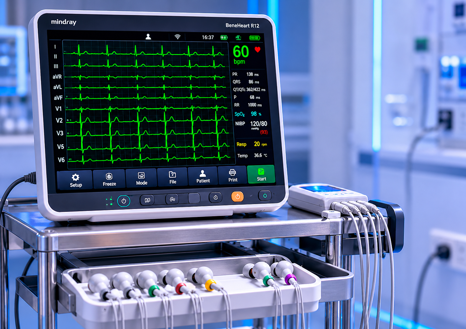
At Jayam Hrudayalaya, our ECG diagnostics are performed with advanced 12-lead digital ECG machines, providing highly accurate readings interpreted by experienced cardiologists.
- Non-invasive & completely painless procedure
- Takes only 5–10 minutes to complete
- Results available immediately
- No special preparation or anaesthesia required
- Safe for all age groups including children & elderly
- Highly accurate when performed by trained technicians
Full Form
Electrocardiogram — "Electro" (electrical) + "Cardio" (heart) + "Gram" (written record)
Also Called
EKG (from German: Elektro-Kardio-Gramm), 12-Lead ECG, Resting ECG
Who Performs?
Trained cardiac technician, after which results are interpreted by a cardiologist
Speed
Paper speed: 25 mm/s | Amplitude: 10 mm/mV is the standard calibration
Electrodes Used
10 electrodes placed on 4 limbs and 6 chest positions to create 12 views
Cost & Access
Affordable, widely available diagnostic test — covered under most insurance & BPL schemes
Every peak and valley on an ECG tracing has a precise medical meaning. Here's what cardiologists look for:
P Wave
Represents atrial depolarization — electrical activation of the upper chambers (atria) of the heart as they contract to push blood into the ventricles.
Q Wave
First negative deflection in the QRS complex. Represents initial depolarization of the interventricular septum. Abnormal Q waves may indicate myocardial infarction.
R Wave
Tallest positive spike in the QRS complex. Represents ventricular muscle depolarization — the main pumping contraction of the heart's lower chambers.
S Wave
Negative deflection after the R wave. Represents the final depolarization of the ventricles — the basal portions of both ventricles.
T Wave
Represents ventricular repolarization — the electrical recovery of the ventricles after contraction, preparing them for the next beat cycle.
U Wave
Small, sometimes visible wave after the T wave. Thought to represent Purkinje fibre repolarization. Prominent U waves may indicate hypokalaemia.
Different clinical situations call for different ECG modalities. Your cardiologist will recommend the most appropriate type based on your symptoms and history.
Resting 12-Lead ECG
The standard ECG performed while you lie still and relaxed. It is the most common type and provides a comprehensive 12-view snapshot of your heart's electrical activity.
- Duration: 5–10 minutes
- Best for: routine screening, chest pain workup
- No preparation needed
- Immediate printed report
- Gold standard for MI diagnosis
Holter Monitor (24–72 hr)
A portable ECG device worn continuously for 24–72 hours to capture intermittent arrhythmias that may not appear during a short resting ECG.
- Duration: 24, 48, or 72 hours
- Best for: palpitations, unexplained syncope
- Patient keeps an activity diary
- Detects paroxysmal arrhythmias
- No activity restriction required
Stress / Exercise ECG (TMT)
Treadmill Test (TMT) — ECG monitored while you exercise on a treadmill or stationary cycle. Reveals abnormalities that only appear during physical exertion.
- Duration: 15–30 minutes
- Best for: exertional chest pain, CAD evaluation
- Fasting for 3–4 hours before test
- Comfortable footwear required
- Physician present throughout
Your doctor may order an ECG in the following situations:
Chest Pain or Discomfort
To rule out or confirm heart attack or angina
Palpitations
Irregular, rapid, or skipped heartbeats felt by the patient
Shortness of Breath
Sudden or unexplained breathlessness at rest or exertion
Dizziness / Fainting
Syncopal episodes or near-blackout spells
Pre-operative Assessment
Mandatory cardiac clearance before any surgical procedure
Medication Monitoring
To monitor cardiac effects of certain drugs (digoxin, antiarrhythmics)
Routine Health Check-up
Preventive screening, especially for patients above 40 years
Electrolyte Imbalance
Changes in potassium, calcium levels affect ECG significantly
Standard intervals, durations and their clinical significance:
| Parameter | Normal Range | What It Measures | Abnormality Significance |
|---|---|---|---|
| Heart Rate | 60–100 bpm | Beats per minute of ventricular contractions | <60 = Bradycardia >100 = Tachycardia |
| P Wave Duration | ≤120 ms | Time for atrial depolarization | Wide P = P-mitrale (LA enlargement) |
| PR Interval | 120–200 ms | AV node conduction time | >200 ms = Heart block <120 ms = WPW |
| QRS Duration | <120 ms | Ventricular depolarization time | Wide QRS = Bundle branch block / VT |
| QT Interval | 350–440 ms | Total ventricular activity (depol + repol) | Long QT = Torsades risk |
| ST Segment | Isoelectric (flat) | Ventricular repolarisation plateau | Elevation = MI / Pericarditis Depression = Ischaemia |
| T Wave | Upright in most leads | Ventricular repolarization | Inverted = Ischaemia / RVH Peaked = Hyperkalemia |
| QRS Axis | -30° to +90° | Direction of ventricular depolarization | LAD = LVH, LBBB RAD = RVH, PE |
An ECG requires virtually no special preparation. However, following these simple guidelines ensures the best quality recording:
Wear Comfortable Clothing
Wear loose, easily removable clothing so electrodes can be placed on your chest, ankles, and wrists without difficulty.
Avoid Lotions & Oils
Do not apply moisturisers, body lotion, or oils to your chest on the day of the test as they can interfere with electrode adhesion.
Remain Still & Relaxed
Muscle movement creates electrical noise. Lie still, breathe normally, and try to stay relaxed during the recording.
Inform About Medications
Inform the technician and cardiologist about all medications you take, especially heart medications, as these affect the ECG findings.
No Fasting Required
For a routine resting ECG, no fasting is necessary. (For stress ECG/TMT, fast for 3–4 hours before the test.)
Remove Metal Objects
Remove jewellery, watches, and metal accessories before the test to avoid interference with the recording equipment.
Recognising these patterns helps cardiologists diagnose a wide range of cardiac conditions:
ST Elevation MI (STEMI)
ST segment elevation ≥1 mm in ≥2 contiguous leads indicates complete coronary artery occlusion — a cardiac emergency requiring immediate intervention.
Non-ST Elevation MI (NSTEMI)
ST depression or T-wave inversion without elevation. Less acute but still indicates significant myocardial ischaemia requiring urgent evaluation.
Atrial Fibrillation (AF)
Absence of distinct P waves, irregularly irregular rhythm. One of the most common sustained arrhythmias, causing stroke risk if untreated.
Ventricular Tachycardia (VT)
Wide QRS tachycardia (≥120 ms) at rate >100 bpm. Life-threatening arrhythmia requiring immediate treatment or cardioversion.
Left Bundle Branch Block (LBBB)
Wide QRS (>120 ms) with characteristic 'M' pattern in V5/V6. New LBBB with chest pain = STEMI equivalent until proven otherwise.
Complete Heart Block (3rd Degree)
Complete dissociation between P waves and QRS complexes. Atria and ventricles beat independently — requires permanent pacemaker implantation.
Long QT Syndrome
Prolonged QTc (>450 ms men, >460 ms women). Risk of Torsades de Pointes — a dangerous form of VT that can degenerate into VF and sudden death.
Wolff-Parkinson-White (WPW)
Short PR interval + delta wave + wide QRS. An accessory pathway bypasses the AV node, causing SVT and potentially fatal rapid conduction in AF.
Right Ventricular Hypertrophy
Right axis deviation, dominant R in V1, deep S in V5/V6. Occurs in pulmonary hypertension, chronic lung disease, and congenital heart defects.
Get Your ECG Done at Jayam Hrudayalaya
Expert cardiac diagnostics with immediate results, affordable pricing, and BPL scheme benefits available.